 | Info
Sheets |
| | | | | | | | | | | | | | | | | | | | | | | | |
 | Out-
side |
| | | | |
|
| | | | |
Result : Searchterm 'Spin Spin Relaxation' found in 2 terms [ ] and 5 definitions [ ] and 5 definitions [ ], (+ 17 Boolean[ ], (+ 17 Boolean[ ] results ] results
| | previous 11 - 15 (of 24) nextResult Pages : [1] [2] [3 4 5] |  | |  | Searchterm 'Spin Spin Relaxation' was also found in the following service: | | | | |
| |  |
| |
|

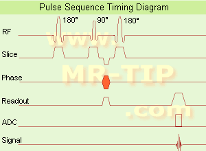
(SE) The most common pulse sequence used in MR imaging is based of the detection of a spin or Hahn echo. It uses 90° radio frequency pulses to excite the magnetization and one or more 180° pulses to refocus the spins to generate signal echoes named spin echoes (SE).
In the pulse sequence timing diagram, the simplest form of a spin echo sequence is illustrated.
The 90° excitation pulse rotates the longitudinal magnetization ( Mz) into the xy-plane and the dephasing of the transverse magnetization (Mxy) starts.
The following application of a 180° refocusing pulse (rotates the magnetization in the x-plane) generates signal echoes. The purpose of the 180° pulse is to rephase the spins, causing them to regain coherence and thereby to recover transverse magnetization, producing a spin echo.
The recovery of the z-magnetization occurs with the T1 relaxation time and typically at a much slower rate than the T2-decay, because in general T1 is greater than T2 for living tissues and is in the range of 100-2000 ms.
The SE pulse sequence was devised in the early days of NMR days by Carr and Purcell and exists now in many forms: the multi echo pulse sequence using single or multislice acquisition, the fast spin echo (FSE/TSE) pulse sequence, echo planar imaging (EPI) pulse sequence and the gradient and spin echo (GRASE) pulse sequence;; all are basically spin echo sequences.
In the simplest form of SE imaging, the pulse sequence has to be repeated as many times as the image has lines. Contrast values:
PD weighted: Short TE (20 ms) and long TR.
T1 weighted: Short TE (10-20 ms) and short TR (300-600 ms)
T2 weighted: Long TE (greater than 60 ms) and long TR (greater than 1600 ms)
With spin echo imaging no T2* occurs, caused by the 180° refocusing pulse. For this reason, spin echo sequences are more robust against e.g., susceptibility artifacts than gradient echo sequences.
See also Pulse Sequence Timing Diagram to find a description of the components.
| | | | | | | | | | | | |  Further Reading: Further Reading: | | Basics:
|
|
News & More:
| |
| |
| | | | | |
| |
|
•
In the 1930's, Isidor Isaac Rabi (Columbia University) succeeded in detecting and measuring single states of rotation of atoms and molecules, and in determining the mechanical and magnetic moments of the nuclei.
•
Felix Bloch (Stanford University) and Edward Purcell (Harvard University) developed instruments, which could measure the magnetic resonance in bulk material such as liquids and solids. (Both honored with the Nobel Prize for Physics in 1952.) [The birth of the NMR spectroscopy]
•
In the early 70's, Raymond Damadian (State University of New York) demonstrated with his NMR device, that there are different T1 relaxation times between normal and abnormal tissues of the same type, as well as between different types of normal tissues.
•
In 1973, Paul Lauterbur (State University of New York) described a new imaging technique that he termed Zeugmatography. By utilizing gradients in the magnetic field, this technique was able to produce a two-dimensional image (back-projection). (Through analysis of the characteristics of the emitted radio waves, their origin could be determined.) Peter Mansfield further developed the utilization of gradients in the magnetic field and the mathematically analysis of these signals for a more useful imaging technique. (Paul C Lauterbur and Peter Mansfield were awarded with the 2003 Nobel Prize in Medicine.)
•
1977/78: First images could be presented.
A cross section through a finger by Peter Mansfield and Andrew A. Maudsley.
Peter Mansfield also could present the first image through the abdomen.
•
In 1977, Raymond Damadian completed (after 7 years) the first MR scanner (Indomitable). In 1978, he founded the FONAR Corporation, which manufactured the first commercial MRI scanner in 1980. Fonar went public in 1981.
•
1981: Schering submitted a patent application for Gd-DTPA dimeglumine.
•
1982: The first 'magnetization-transfer' imaging by Robert N. Muller.
•
In 1983, Toshiba obtained approval from the Ministry of Health and Welfare in Japan for the first commercial MRI system.
•
1986: Jürgen Hennig, A. Nauerth, and Hartmut Friedburg (University of Freiburg) introduced RARE (rapid acquisition with relaxation enhancement) imaging. Axel Haase, Jens Frahm, Dieter Matthaei, Wolfgang Haenicke, and Dietmar K. Merboldt (Max-Planck-Institute, Göttingen) developed the FLASH ( fast low angle shot) sequence.
•
1988: Schering's MAGNEVIST gets its first approval by the FDA.
•
In 1991, fMRI was developed independently by the University of Minnesota's Center for Magnetic Resonance Research (CMRR) and Massachusetts General Hospital's (MGH) MR Center.
•
From 1992 to 1997 Fonar was paid for the infringement of it's patents from 'nearly every one of its competitors in the MRI industry including giant multi-nationals as Toshiba, Siemens, Shimadzu, Philips and GE'.
| | | | | |
• View the DATABASE results for 'MRI History' (6).
| | |
• View the NEWS results for 'MRI History' (1).
| | | | | | Further Reading: | | Basics:
|
|
News & More:
| |
| |
| | | | | |
| |
|
(IR) The inversion recovery pulse sequence produces signals, which represent the longitudinal magnetization existing after the application of a 180° radio frequency pulse that rotates the magnetization Mz into the negative plane. After an inversion time (TI - time between the starting 180° pulse and the following 90° pulse), a further 90° RF pulse tilts some or all of the z-magnetization into the xy-plane, where the signal is usually rephased with a 180° pulse as in the spin echo sequence. During the initial time period, various tissues relax with their intrinsic T1 relaxation time.
In the pulse sequence timing diagram, the basic inversion recovery sequence is illustrated. The 180° inversion pulse is attached prior to the 90° excitation pulse of a spin echo acquisition.
See also the Pulse Sequence Timing Diagram. There you will find a description of the components.
The inversion recovery sequence has the advantage, that it can provide very strong contrast between tissues having different T1 relaxation times or to suppress tissues like fluid or fat.
But the disadvantage is, that the additional inversion radio frequency RF pulse makes this sequence less time efficient than the other pulse sequences.
Contrast values:
PD weighted: TE: 10-20 ms, TR: 2000 ms, TI: 1800 ms
T1 weighted: TE: 10-20 ms, TR: 2000 ms, TI: 400-800 ms
T2 weighted: TE: 70 ms, TR: 2000 ms, TI: 400-800 ms
See also Inversion Recovery, Short T1 Inversion Recovery, Fluid Attenuation Inversion Recovery, and Acronyms for 'Inversion Recovery Sequence' from different manufacturers. | | | | | |
• View the DATABASE results for 'Inversion Recovery Sequence' (8).
| | | | | | Further Reading: | Basics:
|
|
News & More:
| |
| |
| | | Searchterm 'Spin Spin Relaxation' was also found in the following service: | | | | |
| | |
| |
|
Contrast enhanced MRI is a commonly used procedure in magnetic resonance imaging. The need to more accurately characterize different types of lesions and to detect all malignant lesions is the main reason for the use of intravenous contrast agents.
Some methods are available to improve the contrast of different tissues. The focus of dynamic contrast enhanced MRI (DCE-MRI) is on contrast kinetics with demands for spatial resolution dependent on the application. DCE- MR imaging is used for diagnosis of cancer (see also liver imaging, abdominal imaging, breast MRI, dynamic scanning) as well as for diagnosis of cardiac infarction (see perfusion imaging, cardiac MRI). Quantitative DCE-MRI requires special data acquisition techniques and analysis software.
Contrast enhanced magnetic resonance angiography (CE-MRA) allows the visualization of vessels and the temporal resolution provides a separation of arteries and veins. These methods share the need for acquisition methods with high temporal and spatial resolution.
Double contrast administration (combined contrast enhanced (CCE) MRI) uses two contrast agents with complementary mechanisms e.g., superparamagnetic iron oxide to darken the background liver and gadolinium to brighten the vessels. A variety of different categories of contrast agents are currently available for clinical use.
Reasons for the use of contrast agents in MRI scans are:
•
Relaxation characteristics of normal and pathologic tissues are not always different enough to produce obvious differences in signal intensity.
•
Pathology that is sometimes occult on unenhanced images becomes obvious in the presence of contrast.
•
Enhancement significantly increases MRI sensitivity.
•
In addition to improving delineation between normal and abnormal tissues, the pattern of contrast enhancement can improve diagnostic specificity by facilitating characterization of the lesion(s) in question.
•
Contrast can yield physiologic and functional information in addition to lesion delineation.
Common Indications:
Brain MRI : Preoperative/pretreatment evaluation and postoperative evaluation of brain tumor therapy, CNS infections, noninfectious inflammatory disease and meningeal disease.
Spine MRI : Infection/inflammatory disease, primary tumors, drop metastases, initial evaluation of syrinx, postoperative evaluation of the lumbar spine: disk vs. scar.
Breast MRI : Detection of breast cancer in case of dense breasts, implants, malignant lymph nodes, or scarring after treatment for breast cancer, diagnosis of a suspicious breast lesion in order to avoid biopsy.
For Ultrasound Imaging (USI) see Contrast Enhanced Ultrasound at Medical-Ultrasound-Imaging.com.
See also Blood Pool Agents, Myocardial Late Enhancement, Cardiovascular Imaging, Contrast Enhanced MR Venography, Contrast Resolution, Dynamic Scanning, Lung Imaging, Hepatobiliary Contrast Agents, Contrast Medium and MRI Guided Biopsy. | | | | | | | | | | |
• View the DATABASE results for 'Contrast Enhanced MRI' (14).
| | |
• View the NEWS results for 'Contrast Enhanced MRI' (8).
| | | | | | Further Reading: | Basics:
|
|
News & More:
|  |
FDA Approves Gadopiclenol for Contrast-Enhanced Magnetic Resonance Imaging
Tuesday, 27 September 2022 by www.pharmacytimes.com | | |
Effect of gadolinium-based contrast agent on breast diffusion-tensor imaging
Thursday, 6 August 2020 by www.eurekalert.org | | |
Artificial Intelligence Processes Provide Solutions to Gadolinium Retention Concerns
Thursday, 30 January 2020 by www.itnonline.com | | |
Accuracy of Unenhanced MRI in the Detection of New Brain Lesions in Multiple Sclerosis
Tuesday, 12 March 2019 by pubs.rsna.org | | |
The Effects of Breathing Motion on DCE-MRI Images: Phantom Studies Simulating Respiratory Motion to Compare CAIPIRINHA-VIBE, Radial-VIBE, and Conventional VIBE
Tuesday, 7 February 2017 by www.kjronline.org | | |
Novel Imaging Technique Improves Prostate Cancer Detection
Tuesday, 6 January 2015 by health.ucsd.edu | | |
New oxygen-enhanced MRI scan 'helps identify most dangerous tumours'
Thursday, 10 December 2015 by www.dailymail.co.uk | | |
All-organic MRI Contrast Agent Tested In Mice
Monday, 24 September 2012 by cen.acs.org | | |
A groundbreaking new graphene-based MRI contrast agent
Friday, 8 June 2012 by www.nanowerk.com |
|
| |
| | | | | |
| |
|
| | | | | | | |
• View the DATABASE results for 'Lung Imaging' (7).
| | |
• View the NEWS results for 'Lung Imaging' (3).
| | | | | | Further Reading: | Basics:
|
|
News & More:
| |
Chest MRI a viable alternative to chest CT in COVID-19 pneumonia follow-up
Monday, 21 September 2020 by www.healthimaging.com | | |
CT Imaging Features of 2019 Novel Corona virus (2019-nCoV)
Tuesday, 4 February 2020 by pubs.rsna.org | | |
Polarean Imaging Phase III Trial Results Point to Potential Improvements in Lung Imaging
Wednesday, 29 January 2020 by www.diagnosticimaging.com | | |
Low Power MRI Helps Image Lungs, Brings Costs Down
Thursday, 10 October 2019 by www.medgadget.com | | |
Chest MRI Using Multivane-XD, a Novel T2-Weighted Free Breathing MR Sequence
Thursday, 11 July 2019 by www.sciencedirect.co | | |
Researchers Review Importance of Non-Invasive Imaging in Diagnosis and Management of PAH
Wednesday, 11 March 2015 by lungdiseasenews.com | | |
New MRI Approach Reveals Bronchiectasis' Key Features Within the Lung
Thursday, 13 November 2014 by lungdiseasenews.com | | |
MRI techniques improve pulmonary embolism detection
Monday, 19 March 2012 by medicalxpress.com |
|
News & More:
| |
| |
| | | | |
| | | |
|
| |
 | Look
Ups |
| |


